Byers, J.A., Brewer, J. W., and Denna, D.W. 1976. Plant growth hormones in pinyon insect galls. Marcellia 39:125-134. pdf

PLANT GROWTH HORMONES IN PINYON INSECT GALLS

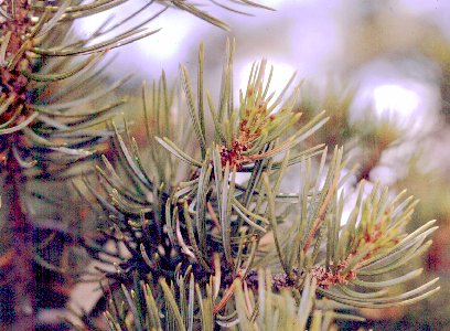
Round galls (redish colored at base) at tips of branches of pinyon pine tree.

Abstract- Larvae of the midge Janetiella sp. near J. coloradensis Felt (Diptera: Cecidomyüdae) cause galls at the base of young needles of pinyon Pinus edulis Engelm. Bioassays of extracts from these galls contained as much as 17 times more auxin activity and as much as 21 times more gibberellin-like activity per needle than extracts from normal needles of the same age. The highest levels of these plant growth substances (per unit volume) occurred during the early stages of gall formation, although abnormally high quantities were found throughout the period of rapid gall growth. Extracts of insect larvae did not contain auxins at detectable levels but traces of substances with gibberellin-like activity were present. Cellular hypertrophy and hyperplasia also support the idea that plant growth controlling substances such as auxins and/or gibberellins probably play an important role in gall formation.

INTRODUCTION

Insect galls of higher plants are generally thought to be caused by the introduction of chemical substances produced by the causative insect (Malpighi, 1675; Plumb, 1953). However, authorities differ as to whether each species of gall-maker releases a different cecidogen (gall inducing compound) or if there is one related group of compounds common to most gall-makers (Boysen-Jensen, 1948; Miles, 1968; Sterling, 1952). Several workers have induced abnormal plant growths by the application of extracts of gall-forming insects (Anders, 1958; Leatherdale, 1955; Lewis & Walton, 1947, 1958; Martin, 1942; Molliard, 1917; Parr, 1939), but what chemicals are involved and cletails of how such growth is controlled remain unresolved. A number of specific inorganic chemicals have been reported to produce gall-like plant growths (Levine, 1950; Mani, 1964). Several amino acids (Anders, 1961; McCalla et al., 1962) as well as adenine containing compounds (McCalla et al., 1962) have also been implicated in insect gall formation.

Plant growth hormones, especially auxins, have been implicated in gall formation because abnormal growths induced by these materials are similar to cells and tissues in natural galls (Arrillaga, 1949; Balch et al., 1964; Brown & Gardner, 1963; Jablonski & Skoog, 1954; McCalla et al., 1962; Schnetzler et al., 1962; Schnetzler et al.,1963; Nitsch,1968; Overbeek, 1966). Most of the evidence for the theory that plant growth hormones are responsible for gall formation comes from work on nematode, fungal or bacterial galls (Bouillenne & Gaspar, 1970) with relatively little work being done on hormone levels in insect-induced galls (Galston & Davies, 1969; Mani, 1964; Newcombe, 1951 ). Quantitative work on these hormones has previously been restricted by lack of appropriately sensitive bioassay or chemical techniques (Burnett & Audus, 1964; Powell, 1964; Sirois,1966,1967; Stahl,1969). The gall midge, Janetiella sp. near J. coloradensis Felt causes a spherical gall (Fig. 1) at the base of Pinyon needles (Pinus edulis Engelm.).

Fig. 1. Deformed Pinus edulis needles caused by Janetiella sp. near J. coloradensis compared with a normal needle.

The female midge lays eggs on terminal buds in June. Feeding by 1 to several larvae during needle development causes the needle base to enlarge abnormally, enveloping the larvae. The gall grows rapidly, reaching 75% of maximum size in about 3 weeks (Fig. 2).

Fig. 2. Growth of Pinus edulis needle galls caused by Janetiella sp. near J. coloradensis showing stage of growth at sampling periods a, b and c.

Larval growth continues until early fall when mature larvae drop to the soil where they overwinter. This gall makes a good experimental system for studying gall formation because growth does not occur until the larva begins to feed, eliminating the possibility of an oviposition fluid initiating gall formation as occurs in some hymenopterous species. In addition, a series of related midge species causes morphologically distinct galls on the same plant (Brewer, 1971; Houseweart & Brewer, 1972), making future comparisons feasible.

In this study we were concerned with determining if auxins and/or gibberellin-like substances occurred in pinyon galls. We also wanted to determine the levels of these materials in normal and abnormal tissue using recently developed extraction, purification, and bioassay techniques. Finally, we wanted to compare abnormal and normal needles to determine what histological and morphological changes occur during gall formation.

MATERIALS AND METHODS


Collection of material Twigs (about 7.5 cm long) containing both galls and normal needles were collected from pinyon trees near Salida, Colorado, on July 23, August 6 and 13, 1972. The plant material was maintained in crushed ice during transit 
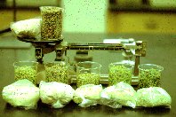
to the laboratory where the normal needles and galls containing the larvae were frozen in liquid nitrogen then each crushed separately with dry ice to a powder-like consistency using a mortar and pestle. The material subsequently was lyophilized and stored at -20oC until needed.

Extraction and purification
Plant material.
Methods used for extraction and purification of plant hormones from pinyon material were modified from techniques of Jones (1968). Although his procedure was primarily for extraction and purification of gibberellic acid (GA), indole-3 acetic acid (IAA) also was found to separate along with gibberellins. We attempted to estimate purification losses of IAA in this process by adding 1000 ug of IAA to a 100 g pinyon needle sample. The sample was then processed and the amount of IAA lost at the various solvent partitioning steps was determined. The results indicated a 2% loss of IAA as determined using the Ehrlich colorimetric reaction (Powell, 1964), but losses of lower levels of IAA, as occurs in natural systems, may not be proportional.

A known amount of the lyophilized pinyon material (59.1 to 79.4 g) from each collection date was homogenized in 125 ml of 80% aqueous methanol with a Waring blender for 3 min at high speed. The subsequent extraction and purification was performed according to the technique of Jones (1968) except that the acidic and basic ethyl acetate extraction steps were reversed chronologically to reduce emulsion formation. The evaporated ethyl acetate extract was redissolved in methanol and then filtered through a 1 cm dia. Whatman No. 1 filter with a syringe equipped with a Swinny adapter.

Thin-layer chromatography and elution of hormones The above filtered methanol extract of plant material was then streaked on 20 X 20 cm silica gel H plates (250 tm thick) with a mechanical streaker and developed in a mixture of benzene, isopropanol, and water (60:20:5 by vol.) at 250oC to a distance of about 14 cm from the origin. After development, each plate was divided into 8 contiguous lateral bands all I.5 cm wide with the first including the origin. Each band of silica gel was scraped from the plates and the materials extracted from pinyon were eluted with methanol into 15 X 150 mm test tubes and evacuated to dryness. Two to 5 tubes containing the dried extract from each band (either gall or normal tissue) were used directly for the auxin and gibberillin bioassays for each date.

Bioassay for auxins
The bioassay for indole growth hormones was modified slightly from that developed by Nitsch & Nitsch (1956) and Sirois (1966, 1967). Oat seed, Avena sativa, of the hulless cultivar `Brighton', was used in all bioassays. The seedlings were harvested when approx. 2 cm tall. The apical 3 mm of each coleoptile was discarded and the next two 6 mm sections below, designated A and B, were placed in beakers of distilled water for 3 h. Five of the coleoptile sections were placed in each 15 X 150 mm test tube containing dried pinyon extract, 1 ml buffer (0.01 M K2HPO4 and 0.005 M citric acid), and 1 ml of water. The test tubes with coleoptile sections were rotated at a 10o angle at 0.5 rev/min for 19 h in the dark at 24oC. The sections were then removed from the tubes and measured for increased length with a dissecting microscope equipped with an ocular micrometer. A standard curve relating coleoptile growth (type A and B) to the concentration of IAA (Fig. 3A) was developed for material from each collection date by following the same procedure but using 1 ml of buffer and various amounts (0-100 ng) of IAA in the test tubes similar to the procedure and results of Sirois (1966, 1967). Corrections for dilutions and sample weight were made for all samples extracted.

Fig. 3. Auxin (IAA equivalents) and gibberellin (GA3 equivalents) standard curves for determining levels in bioassays. Coleoptiles A (C.A) and B (C.B) were plotted separately in the auxin bioassay.

Bioassay of gibberellins
The bioassay of gibberellins was essentially that of Jones & Varner (1967) except for the following modifications: ( 1 ) Naked seed of Hordeum vulgare cv. was used; (2) the barley seeds were weighed in groups of 10 and then surfaced-sterilized with 1.3% sodium hypochlorite for 15 min; (3) the seeds were planted on sterilized germination pads contained in 10 cm Petri dishes in an isolation chamber to reduce fungal contamination; (4) 50 ml Erlenmeyer flasks with 1 ml buffer (0.002 M sodium acetate and 0.02 M CaCl2 ) and 1 ml of various amounts of GA3 (0-10 ng) were used as standards. A unit of a-amylase activity was defined according to Nickells et al. ( 1971 ) as the amount of enzyme able to convert 100 ug of starch to glucose per min. A standard curve of a-amylase release by barley endosperm in response to known quantities of pure GA3 was determined each time pinyon material was extracted and bioassayed. The nanograms of GA3 equivalents in pinyon extracts then were estimated from the standard curves (Fig. 3B). Corrections for dilution, amount of material used, and differences in seed weight were made for all pinyon samples extracted.

Insect material
In order to determine if plant hormones were present in the gall-causing midge larvae, approximately 200 Janetiella sp. near J. coloradensis larvae were dissected from galls and crushed in methanol. The material was tested for the presence of auxins and gibberellin following the same procedure used for the plant material. A non-gall-forming insect species, Tetanops myopaiformis (Otitidae: Diptera), was treated similarly for comparison with the gall-former.

Inhibition
When large quantities of pinyon extract were added to bioassay tubes, growth of the coleoptile sections was inhibited. To test for the level of inhibition, 100 ng of pure IAA was added to bioassay tubes with various amounts of pinyon extract from the August 13, 1972, collection. The larger amounts of pinyon extract caused less growth than would be expected with the larger quantities of IAA. Based on the results of this test, we used 0.3 or 0.5 ml of pinyon extract in most of the bioassay tubes because inhibition was minimal at that level. Although we originally used a correction factor to adjust coleoptile growth for inhibition effects, the corrected values did not differ greatly from those obtained directly and so the latter have been used.

Preparation of thin sections for microscopy
Round galls, and normal needles of the same age were fixed and prepared for sectioning according to the procedures of Sass (1958). After sectioning at 25 um the material was stained with 1% saffranin-0 for 30 min then dehydrated in 95% ethanol and stained with 0.6% acid fast green for 30 sec. Surface areas of cross-sections of the gall or normal needles were determined microscopically using an adjustable ocular grid. Photomicrographs were taken of some of the slides under normal and polarized light.

RESULTS AND DISCUSSION

Auxins in plant tissue
The amount of auxin (IAA equivalents) was substantially higher in galled than normal needles, suggesting that some type of auxin was responsible for gall formation. Galled needles averaged as much as 17 times more auxin than normal needles when calculated on a per needle basis (Fig. 4).

Fig. 4. Auxin levels (IAA equivalents) per needle for normal tissue and gall tissue from Pinus edulis needles.

Average differences in auxin levels calculated on a fresh tissue weight basis were lower although galled tissue still contained as much as 3.7 times more auxin than normal tissue (Fig. 5).

Fig. 5. Auxin levels (IAA) equivalents) per gram fresh weight of normal tissue and gall tissue from Pinus edulis needles.

Differences in the 2 methods of comparison (needle vs tissue weight) are related to the fact that galled needles are heavier than normal ones so fewer are required for a given sample weight. The total amount of auxin may not necessarily determine the amount of galling since an increased concentration of this hormone only in certain cellular organelles or only at certain plant growth stages may effect gall formation. Therefore it is possible that the growth of the gall may actually decrease the total concentration of hormones on a tissue weight basis by causing an increase in weight of plant tissue. This appears to be the case in our study. When quantities of auxin are adjusted to account for varying gall size (mean IAA/needle) per (mean gall volume) the results show that auxin levels, per unit volume, were highest at 12.3 pg/mm3 in the youngest galls collected on July 23 and then dropped to 43 pg/mm3 for the August 6 and August 13 collection dates. In the thin layer chromatographic studies silica gel bands Nos. 4-8 (from the origin) of pinyon extracts were tested for the presence of auxins using the bioassay method described previously. These bands were used because the Rf values were fairly close to IAA. Only eluates from bands 5 and 6 (Rf 0.5 to 0.75) caused increased coleoptile growth as compared to the control. When pure IAA was chromatographed with pinyon extracts it was found to migrate to band 6 (Rf 6.3) with substantial tailing in band 5. It appears, therefore, that part of the material extracted from pinyon galls causes increased coleoptile growth and has extraction characteristics and Rf values in the general range of IAA.

Auxins in Insect larvae
No auxin activity was detected in any of the insect material tested. Any auxin produced by the insect would probably originate in the salivary glands and be released during feeding. Therefore, large quantities need not be present in the insect at any one time but could be continually released and accumulated in the plant tissue. This possibility is supported by the work of Kaldewey (1965) who found that cynipid larvae from oak galls contained 0.5 ng IAA/larva. Ten times that amount/larva was released into agar during a 5-day period following excision from the gall. It is also possible, however, since we found no detectable auxin activity, that the pinyon gall former somehow induces the plant to produce abnormally high auxin levels and thus causes gall formation directly.

Gibberellin-like substances in plant tissue
Higher levels of gibberellin-like materials (GA3 equivalents) were found in galled than normal needles of the same age. Galled needles contained as much as 21 times more gibberellin-like activity than normal needles when calculated on a per needle basis (Fig. 6).

Fig. 6. Gibberellin levels (GA3 equivalents) per needle for normal tissue and gall tissue from Pinus edulis needles.

When levels were calculated on a tissue weight basis, differences were lower although galled tissues still contained as high as 4.8 times as much gibberellin-like activity as normal tissues (Fig. 7).

Fig. 7. Gibberellin levels (GA3 equivalents) per gram fresh weight of normal tissue and gall tissue from Pinus edulis needles.

Differences in the 2 methods of comparison are a result of the tissue weight problems noted for auxin measurements. As with the auxins, when quantities of GA are adjusted for varying gall size (mean GA/needle)/(mean gall vol.) the results show that the highest levels of GA per unit vol. occur in the youngest galls and declines as the galls increase in size. GA levels were 0.17 pg/mm3 on July 23, 0.11 pg/mm3 on August 6 and 0.08 pg/mm3 on August 13. Silica gel band 2 (Rf 0.15 to 0.25 ) had the highest gibberellin activity from chromatographed pinyon extracts. Pure GA3 chromatographed with the pinyon extract migrated to band 2. However, in tests of extracts this band was relatively wide ( 1.5 cm) and may also have contained other of the 40 or more known gibberellins. The presence of other gibberellins could alter the levels calculated since our determinations were based on GA3 equivalents and activity of some of the other gibberellins varies. However, the relative amounts of GA calculated for galled and normal tissue should still be accurate, therefore, the higher levels of activity in galled tissue suggests that gibberillin-like materials probably influence gall formation.

Gibberellin in insect larvae
Bioassays of larval extracts for gibberellin activity indicated a gibberellin level of about 0.07 ng from 204 larvae. This relatively low level of gibberellin, however, only accounts for about 10% of the activity found in galled tissue. It is possible that the gibberellin-like material was introduced with continued salivary secretions into the plant where it accumulated. It may be, however, that the gibberellin activity found in the bioassay was actually the insect moulting hormone, ecdysone (Carlisle et al., 1963) since extracts from a non-gall forming insect species (T. myopaiformis) of similar weight also produced about 0.02 ng of gibberellin-like activity.

The mechanism by which cecidomyii larvae induce or produce higher levels of gibberellin in these galls is unknown and can only be speculated. However, high levels of exogenous gibberellin are known to increase auxin levels in plants (Kuraishi & Muir, 1964; Sastry & Muir, 1953) possibly by increasing the orthophenols which inhibit peroxidase (Kogl & Elema, 1960). Thus, the gall-forming larvae might secrete or induce the production of gibberellins which would in turn increase auxin levels or perhaps act synergistically with IAA to cause the abnormal growths.

Needle morphology
High levels of auxin and/or gibberellin will cause cell expansion (hypertrophy) and cell division (hyperplasia) in many plants (Sachs, 1961; Jablonski & Skoog, 1954; Nitsch,1968). The mesophyll parenchyma cells of galled needles show a remarkable hypertrophy when compared to cells of young needles (Fig. 8A and B).

The average mesophyll parenchyma cell in the round gall increased 13 times in volume and divided 1.5 times more often than a cell from a young needle (Table 1). The results, however, were calculated assuming a spherical cell which only approximates the actual cell shape. There also appeared to be an alteration of plastids and the chloroplasts may have undergone some type of degradation involving the loss of chlorophyll. However the latter may have been an artifact of the staining technique.
Table 1. Comparison of mean cell size and number in cross sections through the middle of normal and galled needles of the same age. (Aug. 1,1972)
Normal needleGalled needle
No. epidermal cells151300
No. cells inside endodermis402501
No. endodermal cells 27not recognizable
No. cells outside endodermis 5581,527
No. cells total9872,028
Area outside endodermis (mm)2 0.548.29
Diameter of average cell outside endodermis (mm)3.25 x 10-28.31 x 10-2
Volume of average cell outside endodermis (mm)32.30 x 10-530.10 x 10⁻⁵
X-fold increase. in volume of average cell outside endodermis113.1

The stunting of the needle length (from 3/4 to 1/2 of normal) during gall formation is partly due to transverse growth of cells as opposed to longitudinal growth. Burg & Burg ( 1966) have reported that auxin interacting with ethylene promotes transverse growth of cells. Therefore, the high levels of auxin found in these galls may possibly induce ethylene production and help stunt needle elongation.

The cellulose content of epidermal cell walls was found to decrease in round galls as determined by inspection of cross sections with a polarizing microscope (Stamm, 1964). Auxin has been reported to induce several polysaccharidases such as (B-1,3 glucanase and exogalactanases (Cleland, 1971 ). These hydrolase enzymes could weaken the cell walls, allowing expansion by decreasing the amounts and lengths of cellulose. Thus, some of the increase in cell size may have resulted from an unfolding of the inner ridges of normal cells. The cellular changes in round galls can be explained by increased levels of auxin and gibberellin. Cytokinins also may be involved in allowing cell hyperplasia to occur but auxin alone will cause the cell to automatically divide once it becomes too large (Jablonski & Skoog,1954).

Numerous starch grains, probably within chloroplasts, were found throughout the mesophyll of the round gall but were absent in young needles. The starch grains were easily observed due to an `x' interference pattern in polarized light because of the concentric layers of starch (Winton, 1906). This starch may provide a rich source of food for the midge larvae. However, Schnetzler et al. (1962), working on a cynipid gall of oak found that when IAA was applied to forming galls from which the larvae were removed, the starch reserves disappeared while the gall continued to enlarge. Gibberellin acid in barley seeds causes starch breakdown but in pinyon needles the physiological effects are probably different.

Auxins and gibberellin-like substances were found to increase dramatically as the normal needle begins to form a gall. In the still-forming gall tissue there was about as much as 17 times more auxin and 21 times more gibberellin-like substances per gall than per normal needle. Calculated on a weight basis, gall tissue had from 2.2 to 3.7 more IAA equivalents and from 2.4 to 4.8 times more GA3 equivalents than normal tissue. The quantities of these growth hormones are not known with certainty to be physiologically active in pinyon gall formation although the amounts are within the range of quantities found in other plant tissues (Overbeek, 1966). If the gibberellin-like substances and auxin are increased due to secondary response from gall formation, they still could affect the morphogenesis of the gall.

This study indicates that the cecidomyiid larvae probably do not secrete auxin, but may secrete gibberellins. (However, the gibberellin activity found in larval extracts, may have been the insect molting hormone, ecdysone, producing gibberellin-like activity in the bioassay.)

These needle galls senesce and fall off the pinyon tree during the winter following gall growth. Most needles remain living on the tree from 4 to 9 years; therefore auxin and/or gibberellin may induce seneseent changes, or change the physiology of the gall to the extent that death results. Auxin apparently has been shown to induce ethylene production in several plant tissues and to alter senescence and abscission (Abeles & Rubinstein, 1964; Burg & Burg, 1966; Morgan & Hall, 1962). Therefore, auxin could cause changes in cell growth and consequently gall formation and later induce ethylene production and senescence. The transverse gall growth may also be aided by ethylene. Cellular hypertrophy and hyperplasia in the gall possibly support the theory that auxin and gibberellin=like materials cause gall formation. Round gall cells increase in volume at least 13 fold from normal needle cells. The amount of cellulose in epidermal cells and the osmotic concentration of cellular contents decreased in round galls showing classical effects of auxin and gibberellin. Other cecidomyüd pinyon galls such as the spindle gall (Houseweart & Brewer, 1972) are probably formed in much the same way as the round gall, since the causative agents are closely related. However, different hormone concentrations and different times of hormone inducement may cause the different morphological galls. This work should be extended to pine tissue culture to determine if the extracts we showed to contain plant growth promoting substances in the bioassay are actually active on pine tissues. Moreover the possible role of cytokinins in needle gall forntations should be studied.

Acknowledgement--The illustrations were prepared by Carol Mortensen, 2420 SW Pickford, Corvallis, Oregon 97330, U.S.A.

REFERENCES

ABELES F. B. & RUBINSTEIN B. (1964). Regulation of ethylene evolution and leaf abscission by auxin. Plant Physiol. 39, 963-969.

ANDERS F. (1958). Aminosauren als gallenerregende Stoffe der Reblaus (Viteus (phylloxera) vitifolii Shimer). Experientia 14, 62-63.

ANDEKS F. (1961). Untersuchungen izber das cedidogene Prinzip der Reblaus ( Viteus (=Phylloxera) vitifolii Shimer) - I. Untersuchungen an der Reblausgalle. Biol. Zbl. 79, 47-58.

ARRILLAGA J. G. (1949). Formation of galls in stems and leaves of sugar cane in response to injections of growth-regulating substances. Phytopathology 39, 489-493.

BALCH R. E., CLARK J. & BONGA J. M. (1964). Hormonal action in production of tumors and compression wood by an aphid. Nature 202, 721-722.

BOUILLENNE C. & GASI'AK T. (1970). Auxin catabolism and inhibitors in normal and crown gall tissues of Impatiens balsamina. Can. J. Bot. 48, 1159-1163.

BOYSEN-JENSEN P. (1948). Formation of galls by Mikiola fagi. Physiol. Plant. 1, 95-108.

BREWER J. W. (1971). Biology of the pinyon stunt needle midge. Ann. ent. Soc. Am. 64, 1099-1102.

BROWN N. A. & GARDNER F. E. (1936). Phytopathological note: galls produced by plant hormones, including a hormone extracted from Bacterium tumefaciens. Phytopathology 26, 708-713.

BUKG S. P. & BURG E. A. (1966). The interaction between auxin and ethylene and its role in plant growth. Proc. natc. Acad. Sci. USA 55, 262-269.

BURNETT D. & AUDUS L. J. (1964). The use of fluorimetry in the estimation of naturally-occurring indoles in plants. Phytochemistry 3, 395-415.

CARLISLE D. B., OSBORNE: D. J., ELLIS P. E. & MOORHOUSE J. E. (1963). Reciprocal effects in insect and plant-growth substances. Nature 200, 1230.

CLELAND R. (1971). Cell wall extension. Ann. Rev. Plant Physiol. 22,197-222.

GALSTON A. W. & DAVIES P. J. (1969). Hormonal regulation in higher plants. Science 163, 1288--1297.

HOUSEWEART M. W. & BREWER J. W. (1972). Biology of a pinyon spindle gall midge (Diptera: Cecidomyüdae). Ann. ent. Soc. Am. 65, 331-336.

JABLONSKI J. R. & SKOOG F. (1954). Cell enlargement and cell division in excised tobacco pith tissue. Physiol. Plant. 7,16-24.

JONES R. L. (1968). Aqueous extraction of gibberellins from pea. Planta 81, 97-105.

JONES R. L. & VARNER J. E. (1967). The bioassay of gibberellins. Planta 72, I55-161.

KALDEWEY H. (1965). Wachstumsregulatoren aus Pf7anzennallen und Larven der Gallenbewohner. Ber. Deutscl: Bot. (Gesell. 78, 73-84.

KOGL F. & ELEMA J. (1960). Wirkungsbeziehungen zwischen lndol-3-essigsäure und Gibberellinsaure. Naturwissenschaften 47, 90.

KUKAISHI S. & MUIR R. M. (1964). The relationship of gibberellin and auxin in plant growth. Plant Cell Physiol. 5, 61-69.

LEATHERDALE D. (1955). Plant hyperplasia induced with a cell-free insect extract. Nature 175, 553-554.

LEVINE M. (1950). The growth of normal plant tissue in vitro as affected by chemical carcinogens and plant growth substances - I. The culture of carrot tap-root meristem. Am. J. Bot. 37, 445-458.

LEWIS I. F. & WALTON L. (1958). Gall-formation on Hamamelis virginiana resulting from material injected by the aphid Hormaphis hamamelidis. Trans. Am. Micros. Soc. 77, 146-200.

LEWIS I. F. & WALTON L. (1947). Initiation of the cone gall of witch hazel. Science 106, 419-420.

MALPIGHI (1675). Anatome plantarum.

MANI M. S. (1964). Ecology of plant galls. Dr. W. Junk, Publishers, The Hague. 434p.

MARTIN J. P. (1942). Stem galls of sugar-cane induced with insect extracts. Science 96, 39.

McCALLA D. R., GENTHE M. K. & HOVANITZ W. (1962). Chemical nature of an insect gall growth- factor. Plant Physiol. 37, 98-103.

MILES P. W. (1968). Insect secretions in plants. Ann. Rev. Phytopath. 6,137-164.

MOLLIARD M. (1917). Production artificielle d'une galle. C.r.hebd. Séanc. Acad. Sci., Paris, 165, 160-162.

MORGAN P W. & HALL W. C. (1962). Effect of 2,4-dichlorophenoxyacetic acid on the production of ethylene by cotton and grain sorghum. Physiol. Plant. 15, 420-427.

NEWCOMB E. H. (1951). Comparative studies of metabolism in insect galls and normal tissues. pp. 417-427. In Plant Growth Substances (Edited by F. Skoog) 1951. University of Wisconsin Press.

NICKELLS M. W., SCHAEFER G. M. & GALSKY A. G. (1971). The action of cyclic-AMP on GA3 controlled responses - I. Induction of barley endösperm protease and acid phosphatase activity by cycfic-3', 5'-adenosine monophosphate. Plant Cell Physiol. 12, 717-725.

NITSCH J. P. & NITSCH C. (1956). Studies on the growth of coleoptile and first internode sections. A new, sensitive, straight-growth test for auxins. Plant Physiol. 31, 94-111.

NITSCH J. P. (1968). Studies on the mode of action of auxins, cytokinins and gibberellins at the subcellular level. pp. 563-580. In Biochemistry and Physiology of Plant Crowth Substances (Edited by F. Wightman & G. Setterfield) 1968. Runge Press, Ltd. Ottawa, Canada. 1642 p.

OVERBEEK J. VAN (1966). Plant hormones and regulators. Science 152, 721-731.

PARR T. J. (1939). Matsucoccus sp., a scale insect injurious to certain pines in the northeast (Hemiptera-Homoptera). J. Econ. Ent. 32, 624-630.

PLUMB G. H. (1953). Formation and development of the Norway Spruce gall caused by Adelges abietis L. Conn. Agric. exp. Sta. Bu11. 566. New Haven, Conn.

POWELL L. E. (1964). Preparation of indole extracts from plants for gas chromatography and spectro-photofluorometry. Plant Physiol. 39, 836-842.

SACHS R. M. (1961). Gibberellin, auxin, and growth retarded effects upon cell division and shoot histogenesis. pp. 42-51. In Papers on Plant Growth and Development. 1967. (Edited by W. M. Laetsch & R. C. Cleland). Little, Brown & Co., Boston. 479 p.

SASS J. E. (1958). Botanical microtechnique. Iowa State Univ. Press. Ames, Iowa.

SASTRY K. K. S. & MUIR R. M. (1953). Gibberellin: effect on diffusible auxin in fruit development. Science 140, 494-495.

SCHNETZLER J. C., MEYER J. & MARESQUELLE H. J. (1962). Observations sur la croissance hiver- nale normale et experimentalement stimulée de la galle lenticulaire de Neuroterus Quercus-baccarum L. = Neuroterus lenticularis 01. C.r. hebd. Séanc. Acad. Sci., Paris, 255, 1643-1645.

SCHNETZLER J. C. (1963). La croissance hivernale de la galle de Neuroterus quercus-baccarum females L. = N. Ienticularis 01.) et sa stimulation par l'hétéroauxine. Marcellia 31, 1-34.

SIROIS J. C. (1966). Studies on growth regulators - I. Improved Avena coleoptile elongation test for auxin. Plant Physiol. 41, 1308-1312.

SIROIS J. C. (1967). A quantitative coleoptile elongation test for growth regulators. pp.1611-1618. In Biochemistry and Physiology of Plant Growth Substances (Edited by F. Wightman & G. Setterfield) 1968. Runge Press Ltd. Ottawa, Canada. 1642 p.

STAHL E. (1969). Thin-layer chromatography. 2nd edn. Springer, New York, 1041 p.

STAMM A. J. (1964). Wood and cellulose science. Ronald Press Co., New York 549 p.

STERLING C. (1952). Ontogeny of the phylloxera gall of grape leaf. Am. J. Bot. 39, 6-15.

SUDSWORTH G. B. (1917). The pine trees of the Rocky Mountain region. U.S.D.A. Bull. No. 460, 1-46.

WINTON E. (1906). The microscopy of vegetable foods. Wiley, New York. 701 p.

home page